The lower limb, in terms of anatomy, is rarely of interest to people with little knowledge in this area. An ordinary person most often represents the leg as a single array of soft tissues that surrounds some large bones. The knee remains the only accessible area for understanding - but its study is usually limited to external landmarks. Most people from all the structures of this joint are called only the patella.
Therefore, it is necessary to dwell in more detail on the issue of the anatomy of the lower limb - more precisely, its section, which includes the thigh and lower leg. It is important not only to determine their exact boundaries, but also to understand the internal structure. This part of the leg is only outwardly unremarkable - it contains the largest anatomical formations in the body.
And all of them are located on the thigh, which is the most important supporting structure of the body. This list includes both skeletal elements and soft tissues - the femur, the sciatic nerve, and the great saphenous vein. But these formations are not isolated - on the thigh and lower leg they are a single whole, differing only in size. Therefore, large sections of the lower limb should be considered as an integral structure, only functionally separated by the knee joint.
Hip
This part of the body has the shape of a truncated cone - its top is the knee, and the base smoothly borders on the body. This appearance is due to the structure of soft tissues - the upper segment of the thigh contains a large number of muscles. In the lower section, the muscles already smoothly pass into wide and strong ligaments, as a result of which the volume of the limb decreases.
The thigh, as a part of the body, has clear boundaries, although the average person is unlikely to be able to correctly indicate them. Therefore, you should consider exactly how it is located in relation to the torso and lower leg:
- The upper border is not transverse throughout - in front it passes along the skin inguinal folds, going obliquely down. Laterally, the leg is delimited from the body along a line drawn through the iliac crest. Behind, the border acquires a transverse direction, passing in the gluteal fold. Its general internal direction corresponds to the plane drawn through the hip joint.
- The lower border of the thigh does not have such structural features, and is calculated quite simply - in relation to the patella. The upper pole of the patella is determined, after which a perpendicular line is drawn 5 centimeters above it.
Knowing the correct boundaries of any part of the body allows the doctor to accurately assess the localization of pathological processes, and also helps to easily find large vessels or nerves in their projection.
Skeleton
The entire static and functional load in this part of the body is assumed by a single bone - the femur. It is the largest indivisible structure of the musculoskeletal system in all respects - size and weight. According to the anatomical classification, the femur has a tubular structure, which is typical for the most loaded and durable formations in the skeleton.
Since it is only one supporting element of the upper segment of the leg, it has to take on the interaction with all soft tissues. Therefore, the femur has a rather interesting structure:
- The upper part consists of the head and neck, which are part of the hip joint. In relation to the segments lying below, they are located at a slight angle. Such a device provides not only good support, but also increases the range of motion in the joint.
- Further, the neck passes into a large tuberous formation - a large and small trochanter of the thigh. They are the site of attachment of the large gluteal muscles.
- Then the largest and longest segment begins - the body of the bone. It has a characteristic tubular structure, expanding slightly in the lower section. On its back surface there is a rough line - a fixation area for some muscles of the thigh.
- The lower section is a rounded extension - it is divided across by a wide depression. These parts are called the condyles and are normally covered by articular cartilage and form the upper half of the knee joint.
The head and neck of the femur have a relatively isolated blood supply, which affects the rate of healing when they are injured.
soft tissues

Between the skin with fatty tissue and the muscle tissue of the upper leg there is another large formation - the fascia lata of the thigh. It is a large connective tissue case that collects all the muscles of the anterior and lateral sections into one large bundle. The durable outer shell gives them the support they need, allowing them to work more efficiently and smoothly.
Inside the muscle bundles there are also tendon septa dividing them into three groups. At the same time, each of them performs a certain amount of movement during contraction:
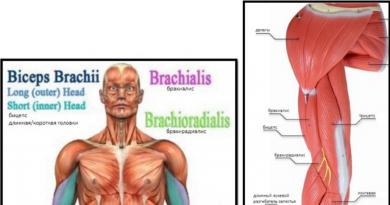
- The front group consists of two long and strong muscles - the tailor and the quadriceps femoris. Their purpose is to carry out flexion of the leg in the hip joint, as well as extension in the knee. The quadriceps muscle in the lower part forms a powerful and wide tendon, which passes through the patella to the lower leg.
- The posterior group is formed by thin and long muscles - the biceps, semimembranosus and semitendinosus muscles. They carry out, on the contrary, extension in the hip joint and flexion in the knee joint. And with fixed legs, their contraction allows you to return the torso from the tilt position.
- The inner group consists of small short muscles - the comb and thin muscles, as well as large, short and long adductors. As a result of their well-coordinated work, the hip is adducted and rotated outward.
The peculiarity of the thigh muscles is their dual purpose - they take on both a powerful static and dynamic load, often combined with each other.
Vessels and nerves

The vast majority of these formations are located in the space located between the anterior and internal muscle groups. Starting from the upper border, the main vascular bundle passes there, providing blood supply to the entire lower limb. The nerves are divided in the opposite way - the largest of them, on the contrary, passes in the back of the thigh.
In general, the arrangement of vessels and nerve bundles is of the main type, characteristic of such a large limb segment. Therefore, they should be considered within these highways:
- Arterial vessels are represented by a large femoral artery, which passes to the limb from the pelvic cavity. It goes in the intermuscular recess along the inner surface of the thigh, giving a deep branch to nourish almost all of the above muscles. The main trunk just above the knee goes deep into the soft tissues, penetrating the popliteal fossa, and going to the lower leg.
- The venous system consists of two parts - the femoral vein represents its deep part, and the large saphenous vein is a superficial vessel. Just below the inguinal fold, they merge, forming a common vein that goes into the cavity of the small pelvis.
- The innervation of the thigh is provided by two systems of nerves located on its opposite sides. Together with the vessels on the inner surface, the femoral nerve emerges. Behind the same passes the most powerful similar structure in the body - the sciatic nerve.
The main type of blood supply and innervation makes the legs vulnerable to injury, since if a vessel or nerve at the level of the thigh is damaged, the entire limb suffers.
Knee-joint

This rather large and complex articulation cannot be ignored - it is both a border and a connecting element between the lower leg and thigh. Therefore, you should consider all the structures included in its composition:
- There are only two main bone elements in the knee joint - these are the condyles of the femur and the articular surface of the tibia. They bear the brunt of the load at rest and during movement.
- But there is also an additional bone - the patella (because of its external shape it is called the patella), which plays an important dynamic role in the joint.
- Inside the joint cavity there are menisci - two semilunar cartilaginous plates that provide tight contact between the articular surfaces of the bones. They also provide good cushioning effect.
- The ligaments complete the entire structure - they surround the knee from all sides, and are even present inside the joint cavity. Their varied position and direction provide the connection with both good strength and mobility.
The points of attachment of the muscles of the lower leg and thigh are located in the areas above or below the knee joint. Despite the fact that they often overlap each other's action, there is no negative effect from this. On the contrary, such a structure ensures the stabilization of the work of all the muscles on the leg among themselves.
Shin
This segment of the lower limb in external and internal structure is very similar to the thigh. The only significant difference is the number of bones that make up their composition. On the lower leg, the supporting structures are represented by two similar elements - the tibia and the fibula. But the essence remains the same - only one of them bears the main load, transferring it to the foot.
The border between the thigh and lower leg does not touch - these structures are completely separated by the knee joint. Therefore, it is necessary to dwell on this issue in more detail:
- The upper border of the lower leg is quite clear - it is a perpendicular plane. It passes through a line drawn 5 centimeters under the lower edge of the patella.
- The lower border has several clear landmarks separating the lower leg from the foot. The most basic and visible even outwardly formations are the ankles. These bony protrusions, located just above the foot, are the final sections of the bones of the lower leg. Their lower pole is the starting point - lines are drawn obliquely upward from it to the front and back surfaces, when connected, they give a clear boundary.
Many people mistakenly refer to the ankles as part of the foot, even though these bony structures are anatomically and functionally an integral part of the lower leg.
Skeleton

The supporting frame of this part of the leg consists of two bones at once, between which the load is still evenly distributed, despite their different sizes. This feature is due to a large number of soft tissues, which completely level out the difference in size to the lower part of the lower leg. Therefore, when moving, the pressure in the lower section is perceived equally by both bones.
Since each of them plays a certain role in the anatomical structure of the lower leg, they differ significantly in structure. Therefore, it is necessary to consider some of their features:
- The tibia occupies an anterior and internal position on the lower leg - it is its contours that protrude through the skin. In the upper part, it has a thickening, forming the lower half of the knee joint. Just below it (under the kneecap) is tuberosity - the place of attachment of the muscles. Then comes the main tubular part, which at the bottom smoothly turns into another thickening - the articular surface and the inner ankle.
- The fibula on the lower leg is located outside, slightly hiding in the upper segment behind a powerful "neighbor". It does not participate in the formation of the knee joint, but is only connected to the tibia with the help of strong ligaments. Then it also passes into the tubular thin part, ending at the bottom with a thickening - the outer ankle.
The ankle is often called a favorite place for fractures - a sharp transition from the narrow part of the bone to the expansion contributes to the development of damage in this area.
soft tissues

All the muscles of the lower leg, as well as on the thigh, are enclosed in strong connective tissue cases, which ensure their isolated work. But due to the small size of the area, they do not cover several muscle groups at once, but only hold separate formations. This feature is due to the connection with the foot - individual muscles provide mobility for both her and the fingers.
For convenience, all the muscles are also divided into three groups, taking into account the position of the cases, as well as their own functions. By this division, they are even more reminiscent of the anatomy of the thigh:
- The most famous among them is the posterior group, which includes the gastrocnemius and soleus muscles of the lower leg. Their fibers are closely adjacent to each other, and when merged in the lower section, they form a powerful Achilles tendon. Functionally with the posterior tibial muscle, as well as the long flexors, they are a single mechanism, providing plantar flexion of the foot and fingers during contraction.
- The anterior group of muscles consists of the tibialis muscle of the same name, as well as the long extensors of the fingers. When contracted, they provide the opposite effect - the dorsiflexion of the foot along with the fingers.
- The most isolated structure is the outer group, which includes the long and short peroneal muscles. Due to their small size, they do not oppose the rest of the muscles, but only carry out an auxiliary and stabilizing effect during their contraction.
The muscles of the lower leg are very unequal in size, therefore, injuries of precisely small muscles that cannot withstand a sharp load are often observed.
Vessels and nerves

The lower leg, unlike the thigh, relatively loses the main type of blood supply and innervation. Starting from the popliteal fossa, there is a rapid division of blood vessels and nerves into several sections, approximately corresponding to muscle cases. Consequently, it is already difficult to single out any large-sized structure in this area:
- A small section of the popliteal artery in the upper segment of the lower leg, leaving the fossa of the same name, quickly divides into two trunks. The first of these is the anterior tibial artery, which passes to the corresponding area through the interosseous membrane. The second branch is the posterior tibial artery, which also gives off a branch to the peroneal muscles.
- The venous system is much more interesting - the deep veins completely correspond in location to the arteries of the same name. But the superficial system includes two formations - the large and small saphenous veins, which merge in the popliteal fossa. The systems communicate with each other via short perforating veins.
- The innervation of the lower leg is provided by bundles of a powerful sciatic nerve - the tibial and common peroneal branches.
Despite the significant separation of the entire vascular and nervous network, the lower leg is still completely dependent on the main location of these pathways on the thigh. Therefore, even their slightest lesion there (especially the nerve) causes a complete loss or decrease in functionality in the underlying departments.