Incredible facts
Some organs in the human body exist not because they serve a purpose, but because they were useful to our ancestors.
They remained as rudiments - physiological remnants of our evolutionary past.
Look closely and you'll see that they make sense within the framework of evolution by natural selection.
The structure of the human body
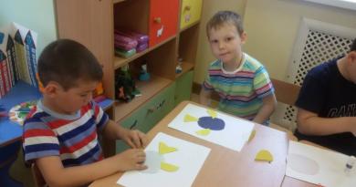
Palmaris longus muscle

The palmaris longus muscle is a vestigial muscle of the forearm. This is a group of muscles that runs from the base of the palm up the arm.
It can be found among many mammals, but it is most developed in those that frequently use their forearms for locomotion, such as monkeys and lemurs. 10-15 percent of people no longer have this muscle, sometimes it is present in only one arm, although this does not affect the compression force.
Often this muscle is removed by surgeons in cases where it is necessary plastic surgery in other parts of the body.
Anterior, superior and posterior auricular muscles

Some people can move their ears. The muscles that allow them to do this are vestigial: the anterior auricularis, superior auricularis and posterior auricularis.
These same muscles allow other mammals to move their ears to better hear sounds and determine their source.

In fact, people try to move their ears in the direction of the sound, but these movements are very small and can be detected using electrodes.
Goosebumps

When we're cold, tiny ones muscles attached to hairs on our body contract and raise the hairs, which causes goosebumps to appear on the skin.
In mammals covered with fur, this creates better insulation and a more intimidating appearance in case of danger. Emotional music can also cause goosebumps because it triggers the fight-or-flight response associated with the release of adrenaline.
Coccyx

The tailbone is also a relic of the tail of our ancestors.
Each of us had a tail at some point in our development - around the fourth week of fetal development. Human embryos are very similar to those of other vertebrates, including the tail. However, in humans and other primates, tail cells are programmed to die.
It is a short fusiform abdomen and a very long tendon, stretching almost from the elbow to the wrist. Oddly enough, 14% of people simply don’t have it.
Anatomy experts say that the absence of this tendon does not affect grip strength in any way. However, in case of any specific injury where the need for transplantation arises, it is good source- a kind of spare part in the human body.
To check if you have it, gather all five fingers into a pinch and bend your wrist - the tendon is clearly visible in the wrist area, provided that it is present. In other mammals, this same tendon is responsible for releasing claws. Apparently, that’s why some people don’t have it - our species doesn’t have the need to extend claws.
Tendons different people may look completely different. The number of tendons and muscles is just as individual.
How to find out something personal about the interlocutor by his appearance

Secrets of “owls” that “larks” don’t know about How does “brainmail” work - transmitting messages from brain to brain via the Internet Why is boredom necessary? “Man Magnet”: How to become more charismatic and attract people to you 25 Quotes That Will Bring Out Your Inner Fighter How to develop self-confidence Is it possible to “cleanse the body of toxins”? 5 Reasons People Will Always Blame the Victim, Not the Criminal, for a Crime Experiment: a man drinks 10 cans of cola a day to prove its harm


- Brachioradialis muscle; m. brachioradialis.
Surface layer
- Extensor carpi ulnaris, m. extensor carpi ulnaris.
- Extensor digitorum, m. extensor digitorum.
- Extensor of the little finger, m. extensor digiti minimi.
The muscles of the forearm, mm.antebrachii, are divided into three groups according to their position: anterior, lateral (radial) and posterior. In this case, the muscles of the anterior and posterior groups are located in several layers. In the anterior group, the muscles lie in four layers.
First (surface layer)
- Pronator teres, m. pronator teres.
- Flexor carpi radialis, m. flехor carpi radialis.
- Palmaris longus muscle, m. palmaris longus.
- Flexor carpi ulnaris, m. flехor carpi ulnaris.
Second layer
- Superficial flexor digitorum, m. flexor digitorum superficialis.
Third layer
- Flexor digitorum profundus, m. flexor digitorum profundus.
- Flexor pollicis longus, m. flexor pollicis longus.
Fourth layer
- Pronator quadratus, m. pronator quadratus
The lateral (radial) group includes:
- Pleradialis muscle; m. brachioradialis.
- Extensor carpi radialis longus, m. extensor carpi radialis longus.
- Extensor carpi radialis brevis, m. extensor carpi radialis brevis.
IN posterior group the muscles lie in two layers.
Deep layer
- Supinator, m.supinator
- Abductor longus muscle thumb brushes, m. abductor pollicis longus.
- Extensor pollicis brevis, m. extensor pollicis brevis.
- Extensor pollicis longus, m. extensor pollicis longus
- Extensor index finger, m. extensor indicis.
Anterior muscle group of the forearm
First (surface) layer
- Pronator teres, m. pronator teres, thick and the most short muscle this layer. It begins with two heads: the larger, humeral head, caput hwnerale, from the epicondylus medialis humeri, septum intermusculare brachii mediale, fascia antebrachii, and the smaller, ulnar head, caput ulnare, originating from the medial edge of the tuberositas ulnae. Both heads form a somewhat flattened abdomen from front to back, which turns into a narrow tendon. The muscle goes obliquely from the inside out and is attached to middle third facies lateralis radii. Action: pronates the forearm and takes part in its flexion. Innervation: n. medianus (C6-C7). Blood supply: muscle branches aa. brachialis, ulnaris, radialis.
- Flexor carpi radialis, m. flexor carpi radialis, bipennate, flat, long muscle. It is located the most lateral of all the forearm flexors. In the proximal part, the muscle is covered only by aponeurosis m. bicipis brachii and m. palmaris longus, and the remaining, large, part of the muscle is covered only by fascia and skin. The muscle begins from the epicondylus medialis humeri, septa intermuscularia and fascia antebrachii and, heading down, passes under the retinaculum flexorum to the base of the palmar surface of the II (III) metacarpal bone. Action: bends and pronates the hand. Innervation: n. medianus [C6-C7-(C8)]. Blood supply: muscle branches a. radialis.
- Palmaris longus muscle, m. palmaris longus, has a short fusiform abdomen and a very long tendon. Lies directly under the skin medially from m. flexor carpi radialis. The muscle originates from the epicondylus medialis humeri, septum intermusculare and fascia antebrachii and, approaching the hand, passes into the wide palmar aponeurosis, aponeurosis palmaris. Action: stretches the palmar aponeurosis and takes part in flexion of the hand. Innervation: n. medianus [(C7) C8].Blood supply: muscular branches of a. radialis.
- Flexor carpi ulnaris, m. flexor carpi ulnaris, occupies the medial edge of the forearm. It has a long muscle belly and a relatively thick tendon.
Starts with two heads:
a) shoulder, caput humerale, from epicondylus medialis humeri and septum intermusculare;
b) ulnar, caput ulnare, from the olecranon, the two upper thirds of the facies dorsalis and the fascia of the forearm.
Heading down, the tendon passes under the retinaculum flexorwn and attaches to the os pisiforme. A number of bundles pass into lig. pisometacarpeum u lig. pisohamatum, which are attached to the hamate and V metacarpal bones. Action: bends the hand and participates in its adduction. Innervation: n. ulnaris (C8, Th1). Blood supply: aa. collaterale, a. brachialis et a. ulnaris.
Second layer
Superficial flexor digitorum, m. flexor digitorum superficialis, covered in front m. palmaris longus and m. flexor carpi radialis, leaving a mark on it in the form of furrows. The muscle itself begins with two heads:
a) humeroulnare, caput humeroulnare. long and narrow, from epicondylus medialis humeri et processus coronoideus ulnae;
b) radial, caput radiale. wide and short, from the proximal part of the palmar surface of the radius.
Both heads, uniting together into a common abdomen, end in 4 long tendons. The latter, moving to the hand, lie in the canalis carpi and are attached to the base of the middle phalanges from the index finger to the little finger. At the level of the proximal phalanges, each tendon is divided into two and therefore is attached not at one, but at two points - along the edges of the base of the middle phalanges. Action: bends the middle phalanges of the fingers from the index to the little finger. Innervation: n. medianus (C7-C8 Th1). Blood supply: aa. radialis et ulnaris.
Third layer
- Flexor digitorum profundus, m. flexor digitorum profundus, is a highly developed, flat and wide abdomen, originating from the proximal half of the facies anterior ulnae and membrana interossea. The muscle is directed downwards, passing into 4 long tendons, which, having passed under the retinaculum flexorum, lie in the canalis carpi, located under the tendons of m. flexor digitorum superficialis. Then each of the tendons m. The flexor digitorum profundus passes between the legs of the tendons of the superficial digital flexor, attaching to the bases of the distal phalanges, from the index finger to the little finger. The tendons of the superficial and deep flexor fingers lie in the common synovial vagina of the flexor fingers of the hand, vagina synovialis communis mm. flexorum digitorum manus. Vaginas of the index, middle and ring finger begin at the level of the head of the metacarpal bones and reach the distal phalanges, without connecting to the common vagina. Only the sheath of the tendons of the little finger is connected to the vagina synovialis communis mm. flexorum digitorum manus. Action: bends the distal phalanges of the fingers from the index to the little finger. Innervation: nn. ulnaris et medianus (C6-C8 Th1). Blood supply: muscle branches a. ulnaris.
- The long flexor pollicis longus, m.flexor pollicis longus, looks like a long single-pinnate flat muscle lying on the lateral edge of the forearm. It starts from the upper 2/3, facies anterior radii and membrana interossea, from epicondylus medialis humeri. The muscle passes into a long tendon, which, moving downwards, lies in the canalis carpi, and is then surrounded by the tendon sheath flexor longus thumb, vagina tendinis m.flexoris pollicis longi, and reaching the distal phalanx, attaches at its base. Action: bends the distal phalanx of the thumb. Innervation: n. medianus (C6-C8). Blood supply: muscle branches aa. radialis, ulnaris et a. interossea anterior.
Fourth layer
The pronator quadratus, m.pronator quadratus, is a thin quadrangular plate of transversely located muscle bundles directly on the membrana interossea. It originates from the distal part of the palmar surface of the ulna and is attached at the same level of the palmar surface of the radius. Action: pronates the forearm. Innervation: n. medianus (C6-C8). Blood supply: a. interossea anterior.
Lateral (radial) muscle group of the forearm
- Brachioradialis muscle, m. brachioradialis, fusiform, occupies the most lateral position. Somewhat below its middle, the muscle passes into a long tendon. It originates from margo lateralis humeri, slightly higher than epicondylus lateralis, and from septum intermusculare brachii laterale. Moving downwards, the muscle attaches to the facies lateralis radii somewhat proximal to the processus styloideus. Action: bends the arm in elbow joint and takes part in both pronation and supination of the radius. Innervation: n. radialis [C5-C6 (C7)]. Blood supply aa. collateralis et recurrens radialis.
- Extensor carpi radialis longus, m. extensor carpi radialis longus, a spindle-shaped muscle with a narrow tendon, significantly longer than the abdomen. In its upper part the muscle is slightly covered by m. brachioradialis, in the distal section the tendon of the muscle is oblique, from top to bottom, intersected by m. abductor pollicis longus and m. extensor pollicis brevis. The muscle starts from the epicondylus lateralis and septum intermusculare brachii laterale, goes down, passes into the tendon, which, passing under the retinaculum ex-tensorum, is attached to the base of the dorsal surface of the os metacarpale II. Action: bends the arm at the elbow joint, extends the hand and takes part in its abduction. Innervation: n. radialis (C5-C7). Blood supply: aa. collaterales (a. profundae brachii) et a. recurrent radialis.
- Extensor carpi radialis brevis, m. extensor carpiradialis brevis, is somewhat covered by the previous muscle in the proximal section, and in the distal section it is intersected by more superficially passing muscles: abductor and extensor pollicis. The muscle originates from epicondylus lateralis humeri, ligg. collaterale and anulare radii. Heading down, it passes into the tendon, which lies next to the tendon of the previous muscle in the sheath of the radial extensor carpi tendon, vagina tendinum mm. extensorum carpi radialium, and is attached to the base of os metacarpale III. Action: extends the hand and abducts it slightly. Innervation: n. radialis [(C5) C6-C7]. Blood supply: aa. collaterales (a. profundae brachii) et a. recurrent radialis.
Posterior muscle group of the forearm
Surface layer
- Extensor carpi ulnaris, m. extensor carpi ulnaris, has a long fusiform abdomen and is located along the inner edge of the dorsal surface of the forearm. The muscle originates from the epicondylus lateralis humeri, margo posterior ulnae and the articular capsule of the elbow joint. Having passed into a short but powerful tendon enclosed in the sheath of the extensor carpi ulnaris tendon, vagina tendinis m. extensoris carpi ulnaris, the muscle is attached to the base of the dorsal surface of os metacarpale V. Action: retracts the hand to the ulnar side and extends it. Innervation: n. radialis [(C6) C7-C8].Blood supply: a. interossea posterior.
- Extensor digitorum, m. extensor digitorum, has a spindle-shaped abdomen, and in the direction of the muscle bundles it is bipinnate. The muscle lies directly under the skin, closer to the lateral edge of the dorsum of the forearm, and borders on the ulnar side with m. extensor carpi ulnaris and with m. extensor digiti minimi, and with radial - with mm. extensores carpi radiales, longus et brevis. The muscle originates from the epicondylus lateralis humeri, the joint capsule of the elbow joint and the fascia of the forearm. In the middle of its length, the muscle belly turns into 4 tendons, which, passing under the retinaculum extensorum, are surrounded, together with the extensor tendon of the index finger, by the sheath of the extensor tendons of the fingers and index finger, vagina tendinum mm. extensoris digitorum et extensoris indicts, reaching approximately the middle of the metacarpal bones. Moving onto the hand, the tendons are connected to each other by non-permanent thin intertendinous joints, connexus intertendinei, and at the base of the proximal phalanx, from the index finger to the little finger, each tendon ends in a tendon extension that fuses with the articular capsule of the metacarpophalangeal joint. Tendon sprains are divided into 3 legs, of which the lateral ones are attached to the base of the distal phalanx, and the middle one is attached to the base of the middle phalanx. Action: straightens the fingers, also taking part in the extension of the hand. Innervation: n. radialis (C6-C8). Blood supply: a. interossea posterior.
- Extensor of the little finger, m. extensor digiti minimi, is a small fusiform abdomen lying directly under the skin in the lower half of the dorsal surface of the forearm, between m. extensor carpi ulnaris and m. extensor digitorum. The muscle starts from epicondylus lateralis humeri, fascia antebrachii and lig. collaterale radiale and, moving downwards, passes into the tendon lying in the vagina of the extensor tendon of the little finger, vagina tendinis m. extensoris digiti minimi. Coming out of the vagina, the tendon connects with the extensor tendon of the fingers, going to the little finger, and is attached with it to the base of the distal phalanx. Action: straightens the little finger. Innervation: n. radialis (C6-C8). Blood supply: a. interossea posterior.
Deep layer
- Arch support, m. supinator, has the appearance of a thin diamond-shaped plate located at the proximal end of the forearm from its outer side back surface. The muscle originates from epicondylus lateralis humeri, crista m. supinatoris ulnae and the articular capsule of the elbow joint, is directed obliquely downwards and outwards, covering the upper end of the radius, and is attached along it from the tuberositas radii to the place of attachment of m. pronator teres. Action: rotates the forearm outward (supinates) and takes part in straightening the arm at the elbow joint. Innervation: n. radialis [(C5) C6-C7 (C8)]. Blood supply: aa. recurrens radialis, recurrens interossea.
- Abductor pollicis longus muscle, m. abductor pollicis longus, has a flattened bipinnate abdomen, turning into a thin long tendon. The muscle lies in the distal half of the dorsolateral surface of the forearm and in its initial part is covered by the m.extensor carpi radialis brevis and m. extensor digitorum, and in the lower section - directly under the fascia anterbrachii and skin. The muscle originates from the posterior surface of the radius and ulna and from the membrana interossea, moving obliquely downwards, bends around the radius with its tendon and, passing under the retinaculum extensorum, attaches to the base of the first metacarpal bone. Action: abducts the thumb, taking part in the abduction of the entire hand. Innervation: n. radialis [C6-C7 (C8)]. Blood supply: aa. interosseae posterior et anterior.
- Extensor pollicis brevis m. extensor pollicis brevis, located in the lower part of the forearm along the lateral edge of its dorsal surface. The muscle starts from the membrana interossea, facies dorsalis radii and crista ulnae, goes obliquely downwards, lying next to the tendon m. abductor pollicis longus. The tendons of these two muscles are surrounded by the sheath of the tendons of the long abductor muscle and the short extensor muscle of the palm of the hand, vagina tendinum mm. abductoris longi et ex-tensoris brevis pollicis. Having passed under the retinaculum extensorum, the muscle is attached to the base of the dorsal surface of the proximal phalanx of the thumb. Action: extends and slightly abducts the proximal phalanx of the thumb. Innervation: n.radialis [C6-C7 (C8)]. Blood supply: aa. interosseae posterior et anterior.
- Extensor pollicis longus, m. extensor Vasa et nn. interossei M. extensor digitorum pollicis longus, has a spindle-shaped abdomen and a long tendon. It lies next to the previous muscle and starts from the membrana interossea, margo interosseus ulnae and facies posterior ulnae and, heading down, passes into the tendon, which lies in the sheath of the tendon of the long extensor of the thumb, vagina tendinis m. extensoris pollicis longi. Then, going around the first metacarpal bone and emerging on its dorsal surface, the tendon reaches the base of the distal phalanx, where it is attached. Action: extends the thumb of the hand and partially abducts it. Innervation: n. radialis [(C6) C7-C8]. Blood supply: aa. interosseae posterior et anterior.
- Extensor index finger, m. extensor indicis, has a narrow, long, fusiform abdomen, located on the dorsal surface of the lower half of the forearm, covered with m. extensor digitorum. Sometimes the muscle is missing. It originates from the lower third of the facies dorsalis ulnae, passes into a tendon that passes under the retinaculun extensorum, and, together with a similar extensor tendon of the digitorum, passing through the synovial sheath, approaches the dorsum of the index finger and is woven into its tendon extension. Action: extends index finger. Innervation: n. radialis [(C6) C7-C8]. Blood supply: aa. interosseae, posterior et anterior.


Ecology of life. Cognitive: 200 muscles are activated with just one step. The heart, the most resilient muscle in the body, works constantly. Muscles grow and train...
200 muscles are activated with just one step. The heart, the most resilient muscle in the body, works constantly. Muscles grow and train; tons of sports literature has been written about them. We will tell you the most interesting things.
1. How many muscles are there in total?
In total, there are from 640 to 850 muscles in the human body. During simple walking, the body uses up to 200 muscles. Muscle tissue is 15% denser and heavier than fat tissue, so a trained person can outweigh an overweight but unathletic person of the same height. Muscles account for an average of 40% of body weight.
2. The very best muscles
The most enduring muscle in humans is the heart, the shortest is the stapedius (it strains the eardrum in the ear). Its length is 1.27 millimeters. Longest muscle human body- tailor's. The most fast muscle- blinking. There are different opinions about which muscle in the body is the strongest. It is often said that the most powerful muscle is the tongue, but the tongue is made up of several muscles, so this point of view is false. Are very strong masticatory muscles(their pressure can reach 100 kilograms), as well as calves and gluteal muscles.
3. Such different muscles
Human muscles are not the same. Therefore, they need to be trained differently, and time for recovery and different groups muscles are different. The triceps recover the fastest, the back muscles the slowest. This must be taken into account when training; muscles need rest no less than load, since the growth of muscle fibers occurs due to the supercompensation effect. Full muscle recovery occurs only 48 hours after intense exercise.
4. Muscle endurance
Endurance is the ability of a muscle to maintain performance over time. The most enduring muscle of the human body, as we have already said, is the heart. According to doctors, the “safety margin” of the average heart is at least 100 years. Muscles begin to tire when they run out of glycogen, which also explains fatigue a large number calcium in muscles. Previously, it was believed that the main culprit of fatigue was lactic acid. A study was conducted at Columbia University in which mice swam daily for three weeks and cyclists trained for three days. It turned out that after physical exercise in the chemical structure of the ryanodine receptor, which is responsible for muscle contraction, there were major changes- a gap appeared in the cell membrane through which calcium leaked into the muscle cells.
5. Muscles and emotions
It is known that the movement of facial muscles is directly related to human emotions. At the beginning of the last century, the Russian scientist Ivan Sikorsky compiled a classification of facial expressions: the muscles around the eyes are responsible for the expression of mental phenomena, the muscles around the mouth are for the expression of acts of will, and all the muscles of the face express feelings. In 2011, scientists were able to discover that human facial expressions arise long before his birth. Even during the prenatal period, the child is already able to move his facial muscles, smile, raise his eyebrows in surprise or frown. Facial muscles make up 25% of the total number of muscles; during a smile, 17 muscle groups are involved, during anger or crying - 43. One of the best ways maintaining smooth skin on the face - kissing. They work from 29 to 34 muscle groups.
6. Muscles and genes
Amazingly, muscle training affects not only the person himself, but also his genes. Modifications occur in them, which subsequently help the muscle fibers to be ready for new loads. In order to prove or disprove this, scientists from Aarhus University recruited a focus group of 20 volunteers and conducted a 20-minute aerobic exercise on an exercise bike. After the study, a biopsy of the subjects' quadriceps was taken to see how the genes had changed in their cells. It turned out that physical activity activates muscle-related genes. This is because cells store DNA using methyl groups. If they are removed, the gene information is converted into enzymes and proteins that are needed to burn calories, gain muscle mass and oxygen consumption. After the experiment, the number of methyl groups decreased in all study participants - that is, the muscles adapted to the increase in metabolism.
7. Muscles and telepathy
A simple person is not able to establish control over all the muscles of the body, therefore unconscious muscle contractions can serve for knowledgeable people as an indicator of hidden thoughts or planned actions. Psychologists high level and "telepaths" can use knowledge about these processes. Wolf Messing, one of the most famous telepaths, explained his phenomenal abilities not by magic, but by a thorough knowledge of the work of human muscles. He said: “This is not mind reading, but, so to speak, “muscle reading”... When a person thinks intensely about something, brain cells transmit impulses to all the muscles of the body.”
8. Palmaris longus
Only one out of six people on earth still have long arms on both arms. palmar muscles. Some people only have them on one of their hands. These muscle fibers are responsible for the release of claws in animals. A person, of course, does not need such a function. The palmaris longus muscle is thus a rudiment used by surgeons, if necessary, as a material for muscle transplantation.
9. Muscles and chocolate
Oddly enough, one of the most healthy products for the heart and for the muscles in general is dark chocolate. Research conducted at Wayne State University in Detroit revealed the effect of the substance epicatechin contained in dark chocolate on the growth of mitochondria in muscle cells. Scientists at the University of L'Aquila also conducted a study in which they gave subjects one hundred grams of chocolate for 15 days and measured their blood pressure. During the experiment, people's blood pressure normalized and their blood circulation improved. Accordingly, moderate consumption of dark chocolate can be considered as a prevention of heart disease and atherosclerosis.
10. Muscle loss
Muscles don't last forever. After 40 years, they begin to be actively burned; a person begins to lose from 2 to 3 percent per year muscle tissue, after 60 years - up to 5%. Therefore, training in adulthood is no less important than in youth. published
4957 0
Tendons are used as tendon autografts, the removal of which does not cause significant functional or cosmetic impairment.
The tendon of the palmaris muscle has a significant length (from 15 to 20 cm or more, including the intramuscular part), a sufficient area cross section and strength. Its loss does not cause functional impairment, and its removal does not cause technical difficulties.
The disadvantages of this source tendon grafts include limited quantity plastic material, absence of tendon in 15% of people and sometimes insufficient length. In this regard, the palmaris longus tendon is most often used for plastic surgery of the flexor tendon on the short fingers of the hand (I and V), when only one finger is damaged. At multiple injuries fingers, it is preferable to use other sources of plastic material.
Taking technique. You can check the presence of the palmaris longus tendon by straining the straightened fingers of the hand while bending it slightly at the wrist joint (Fig. 14.5.1). From a small transverse approach, the tendon is exposed at the junction with the palmar aponeurosis. When doing this, you should be careful not to damage the nearby median nerve.
Rice. 14.5.1. Test to assess the presence of the palmaris longus tendon.
The end of the tendon is sutured and cut off, after which, by pulling the ligatures with simultaneous palpation, it is easy to determine its course under the skin. This allows, from two additional transverse approaches, to completely isolate the tendon to its outer muscle section (Fig. 14.5.2, a), after which it is cut off from the muscle belly. This procedure can be performed from one access using a special instrument - a tendon raspator (Fig. 14.5.2, b).

Rice. 14.5.2. Schematic illustration of the approaches used to harvest the palmaris longus tendon (explanation in text).
Long extensor tendons of the II-V toes. This source is characterized by a significant number of donor tendons (4 on each foot), their significant length (up to 25-30 cm), as well as minor loss of function and cosmetic defect after collection.
At the same time, sometimes the tendons are not thick enough (usually on the 4th-5th fingers), and their isolation over the entire length is technically difficult. This source of tendons is widely used in hand surgery, as well as in reconstructive operations on other segments.
Taking technique. On the bloodless segment, from short (5 mm) transverse incisions at the level of the heads of the metatarsal bones (Fig. 14.5.3, b), the ends of the tendons of the long extensors of the II-V fingers are isolated, sutured and cut off. In this case, the short extensor tendons of the same fingers must be preserved.
From the next transverse approach at the level of the transverse tarsal joint (Shopard's joint), all 4 tendons located next to each other can be isolated. From the third longitudinal access up to 8 cm long, located immediately above the proximal edge of the suspensory extensor tendon ligament, the long extensor tendons of the II-V fingers are exposed, which usually cannot be separated, since they represent one common trunk.
After this, the tendon sheath is carefully opened and a Rozov conductor is inserted into it in the distal direction, trying to pass to the nearest incision along the surface of the extreme tendon. With the help of a guide, each tendon is brought into the proximal wound and, thus, the common tendon trunk is separated. Then the tendons are cut off from the muscle and after removing the tourniquet and stopping the bleeding, the wound is tightly sutured in layers (including the deep fascia).
When using a tendon raspatory, access to the lower legs is not necessary, and the procedure for taking grafts is simplified (Fig. 14.5.3, a).

Rice. 14.5.3. Approaches and stages of harvesting the tendons of the long extensor toes (explanation in the text).
For special indications, extensor toe longus tendons can be incorporated into the dorsalis pedis flap and used as perfused grafts.
The superficial digital flexor tendons are used for plastic surgery of the deep digital flexor tendons. Their advantages include their significant thickness, length and quantity, as well as the simplicity of the picking technique. According to their characteristics, they are best suited for deep tendon replacement. However, their use also has significant negative sides.
First of all, in short-fingered subjects these tendons may be relatively short. This forces them to be taken together with the intramuscular part, after which the muscle can no longer be used, and the strength of flexion of the finger is noticeably reduced. In addition, taking the superficial flexor tendons requires significant access, which is also unprofitable from a cosmetic point of view.
It is important to note that this donor zone is located along the osteofibrous canals of the fingers and therefore is the most unfavorable (in comparison with any other zones) due to the negative influence of scar adhesions that inevitably form around the deep flexor tendons of the donor finger. In the postoperative period, such a finger itself requires full rehabilitation, which may not always result in complete restoration of function.
This is why it is advisable to use the superficial flexor tendons of only the injured finger and only when the level of injury is in the “critical” zone.
If injury occurs at a more proximal level, the tendon graft may become too short to accommodate effective application. Transposition of the superficial flexor tendon from the adjacent, longer and undamaged (!) finger is a gross mistake.
Taking technique. Together with the proximal end of the damaged tendon of the deep digital flexor, the superficial tendon is isolated from the appropriate access on the hand and brought into an S-shaped wound on the forearm. Then the tendon of the superficial flexor of the finger is isolated to the level of the muscle and cut off, having previously sutured its remaining end in the muscle. The latter is sutured to the central end of the deep flexor tendon after its restoration.
The plantaris tendon is of considerable length and thickness, which allows it to be used in hand tendon surgery. Its disadvantage is the limited amount of plastic material, which allows this source to be used only for tendonoplasty on one finger. Additionally, the tendon is missing in 7% of people and cannot be identified before surgery.
Taking technique. The plantaris tendon is isolated from a 5-cm vertical incision anterior to the medial border of the calcaneal tendon and harvested using a tendon rasp (Fig. 14.5.4). In this case, the instrument should pass parallel to the axis of the lower leg when extended in knee joint limbs.

Rice. 14.5.4. Scheme of taking a transplant from the tendon of the plantaris muscle (explanation in the text).
The fascia lata of the femur is a virtually unlimited source of plastic material and, when replacing large tendons, must be rolled into a tube. Due to the fact that its surface does not have such high sliding properties, flaps from the fascia lata of the thigh are not used to replace defects in the flexor tendons of the fingers.
At the same time, their transplantation can give good result when replacing other tendons, including in the form of blood-supplied grafts, including fasciocutaneous flaps from the outer surface
hips.
Tendon autoplasty
The use of autotendons for tendonoplasty is most common in clinical practice. Depending on the specific conditions, five main options are used.One-stage non-vascular graft tendonoplasty is the most common procedure in which a tendon insert is sutured into the tendon defect.
In the vast majority of cases, this type of operation is performed for chronic injuries of the flexor tendons of the fingers.
Two-stage tendonoplasty is used exclusively in surgery of the flexor tendons of the fingers and consists in the fact that during the 1st stage of treatment, more favorable conditions are created for the subsequent transplantation of a tendon graft.
Tendoplasty combined with transplantation of complex skin flaps. When tendon defects are combined with skin defects, these two problems can be solved simultaneously, since only when the tissues surrounding the tendon are in a normal state can their function be restored.
Most often this situation occurs with injuries of the forearm in the lower third. After transplanting a complex skin flap into the defect, tendon grafts can be passed through the latter.
These two tasks can be solved sequentially during two-stage treatment. This significantly lengthens its duration and requires repeated intervention in the same anatomical area.
Transplantation of blood-supplied tendon grafts. When a soft tissue defect is combined with a tendon defect, blood-supplied tissue complexes, including tendons, can be used.
For this purpose, a dorsal foot flap can be used, taken with the tendons of the long extensor muscles of the II-V fingers. Tissue complexes from the outer thigh may include fascia lata, flaps of which can replace tendon defects.
Tendon transposition is one of the methods for replacing tendon defects when a nearby tendon is used, the muscle of which can be switched to a new function without significant functional loss. Most often, one of the paired tendons adjacent to the defect area is used (superficial and deep flexor tendons, common and intrinsic extensors of the II and V fingers).
V.I. Arkhangelsky, V.F. Kirillov