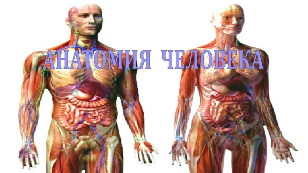
Human hands and feet have their own structural features. They have evolved along with man, adapting to functional needs. Next, we will take a closer look at the anatomical components of the limbs - the joints of the legs and arms, muscles, ligaments, nerve and blood vessels.
Anatomical features
The human legs have a more massive bone and muscle structure, unlike the arms, as they are subjected to significant daily load. After all, their main function is the movement of the body. Therefore, the lower limbs have a number of features - the ability to straighten the knee joint, a powerful ligamentous apparatus, the pelvic part has an expanded shape. This makes it possible to support the internal organs, and the large distance between the joints of the pelvis contributes to the stability of the body.
Human legs are longer than arms. Although in a newborn the length of the lower limbs is only sixty-five percent, but in an adult it is already one hundred and seven. The legs are stronger and stronger, and the arms are capable of a wider range of range in their movements. This is achieved due to the length of the clavicles, the location of the shoulder blades and the features of the shoulder girdle. The joints of the shoulders are slightly moved away from the body due to the collarbone, and due to the connection with the chest, the arms rest on the skeleton of the body.
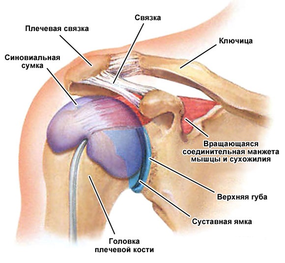
Thanks to the shoulder blades for the upper limbs, freedom of movement is ensured. The shoulders are connected to the body by muscle tissue. The fossa in the joint of the scapula is a quarter of the articular surface of the shoulder bone in size. Thanks to this, the arms move freely, but at the same time, it is at this point that they are highly prone to dislocations.
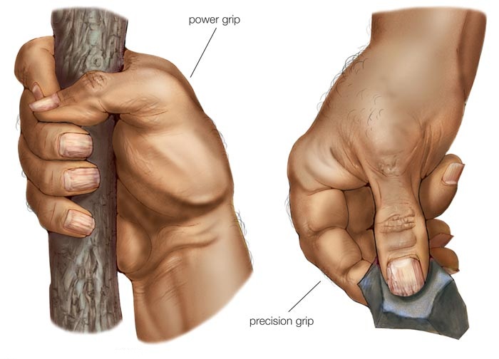
The human forearm is shorter than the humerus. Due to which the hand is capable of quick and precise movements, and also makes it possible to apply less effort to lift heavy objects. In humans, the arm is adapted to perform a wide range of movements due to the predominance of flexors in muscle tissue over extensors, and pronators over supinators.
Role and functions in the body
The limbs of a person perform important functions, thanks to which it is possible to move around, play sports, work, and perform household daily tasks.
The main functions of the upper limbs:
- the shoulder girdle holds the rest of the arm, due to which it can withstand heavy loads;
- the movable part of the limb performs a wide range of different movements due to large joints;
- daily precise movements are performed at the expense of small joints, in the hand, as well as the forearm;
- the fixed component of the limb performs a supporting function for the mobile one, a huge number of muscle fibers help it in this;
- the blood circulation of the hand is provided by the vessels, and the nerve fibers play an innervation role in the movements and reactions of the hand.
The legs carry the entire weight of the body and allow movement. The joints are more enduring than those of the arms, the muscular corset is more powerful. With excessive physical exertion, or excess weight, the cartilage tissue in the joints wears out very much. Thus, the functional ability of the limbs suffers. The location of the joints and the ligamentous apparatus ensures the stability of the body while standing, walking, running.
Among the muscle tissue in the legs there are many oblique fibers, which, due to elongation, provide tirelessness and endurance of the limbs. A distinctive feature of the legs are the feet. They have springy vaults of transverse and longitudinal type. Due to what a person is capable of upright walking.
The feet provide weight distribution on the leg, absorb shocks, points during walking, promote smooth movement and stability. The arch of the foot in a person is not formed immediately at birth, but closer to a year, when the child takes the first steps.
Detailed structure
The main structural units of the limbs are joints, bones, muscles, blood vessels, nerve fibers, ligaments. Separately, it is worth highlighting the hands and feet, as important and complex in structure elements of the limbs. Next, we will take a closer look at each unit of the anatomy of the legs and arms.
Bones
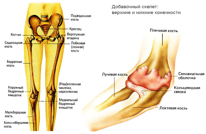
The arm is attached by the collarbone and scapula. The clavicle is located at the top of the sternum, and the scapula is located above the upper ribs from the back. Further, it is connected to the articulation with the bone of the shoulder. The latter holds the rest of the bones of the hand. The next part is called the forearm. It consists of the ulna and radius bones, which are connected to the joint.
The next department is the most complex and multifunctional - the brush. Which in turn is divided into sections:
- wrist - eight bones arranged in two rows. They are part of the wrist joint;
- metacarpal bones - five bones located from the wrist to the phalanges. They almost do not move, as they are supporting for the fingers;
- phalanges of the fingers - four fingers in their structure have proximal, middle and distal bones. And the thumb consists of two - the main and nail bones.
The upper leg consists of the ischium, ilium, and pubis of the pelvis. They are the support for the body. At the age of eighteen, these bones fuse and form the acetabulum. It includes the femur, forming a joint. Thanks to him, the leg rotates and supports the weight of a person.
The femur ends at the patella, which enters the patella. Next is the tibia and the small. And actually the last element is the foot. In its structure, there are twenty-six bones.
leg bones
The foot is divided into three parts, just like the hand:
- tarsus - talus (connects to the lower leg), calcaneus, navicular, lateral, intermediate, medial and cuboid;
- metatarsus - these are five bones that connect the previous section of the foot with the phalanges of the fingers;
- toes - four fingers consist of three phalanges (proximal, middle, distal). The thumb has two bones, the middle one is missing.
joints
The arm consists of three large joints (shoulder, elbow and wrist), as well as a huge number of small ones. Large joints of the upper limbs:
- The shoulder joint is a spherical connection of the shoulder bone with the scapula. Due to the large articular surface of the scapula, this articulation has a large range of motion.
- The elbow joint is formed by three bones (humerus, ulna and radius). It has a block fastening system, due to which it can work for flexion, extension, as well as adduction, abduction.
- The wrist joint - is formed due to the radius and nearby. Movement is possible in three planes.
Shoulder structure
The hand consists of four types of joints:
- mid-carpal - formed between two rows;
- carpometacarpal joints;
- metacarpophalangeal - perform the function of holding the phalanges on the fixed area of \u200b\u200bthe brush;
- interphalangeal - four fingers are formed due to two joints, and the thumb has one.
The longest range of motion is provided by the last two groups of joints. The remaining joints perform an additional function, not particularly participating in movements.
| Articulation name | Short description |
| Hip | The spherical joint is the most powerful in the human body, performs a wide range of movements. The articulation is not palpable by hand, as it is located deep under the muscle tissue. Formed by the head of the femur and the acetabulum. |
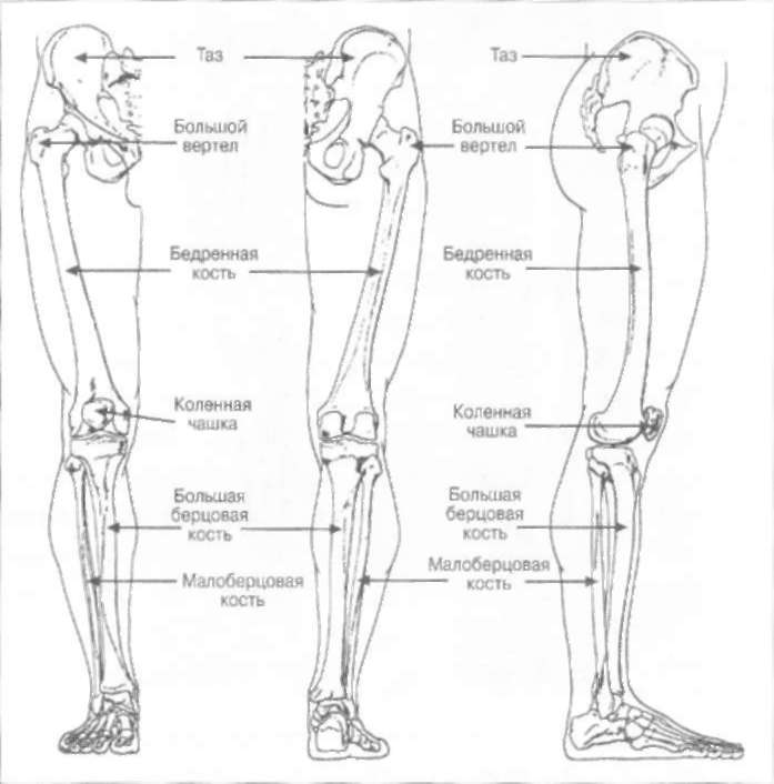
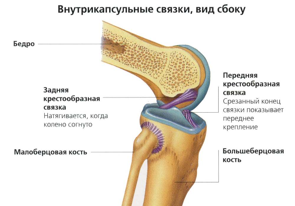
| Knee | Condylar type, formed by three bones - the femur, tibia and patella. Inside there are two lunate partitions made of cartilaginous tissue. They are called menisci. In the joint there are several bags with synovia, not connected to the main cavity of the joint. The knee can flex, extend, and rotate within a small range. Ligaments are placed crosswise. |
| Ankle | Block articulation of the leg and foot. It is formed from two tibia bones (large and small), which are arranged in the form of a fork and capture the talus. The articulation bag resembles a cuff, which has strong walls on the side, and the front and back are represented by loose folds. Functional tasks - flexion and extension. |
| finger joints | The phalanges, like the hands, have two joints on four fingers and one on the thumb. They are less mobile than on the arm, since there is no functional need for this. |
Bundles
Ligaments connect bones to each other, fixing them in a certain position, and limit the amplitude of their movements. Ligaments are located in and around joints, named identically to the bony joints to which they are attached.
In the hands are such bundles:
- three ligaments of the collarbone;
- three articular-humeral (upper, lower and middle);
- four ligaments of the elbow (ulnar, anterior, radial and posterior);
- ligaments in the wrist joint (lateral, radial, ulnar, dorsal, palmar and intercarpal);
- a ligament that closes the flexor retinaculum - a special ligament with a special purpose, which is located in the carpal tunnel, prevents clamping of blood vessels and nerves;
- ligaments of the hand - there are many of them, they connect the small bones of the hand.
The lower limbs are represented by such ligaments:
- - three longitudinal and one circular;
- knee - large and fibular, internal cruciate, popliteal and patellar ligament;
- ankle - external lateral (anterior, posterior and calcaneal fibular), four internal lateral, interosseous (passes through the entire lower leg). There is also a posterior inferior (does not allow bones to turn inward), anterior fibular and transverse.
muscles
The muscles of the leg are very strong, since their main function is to ensure the mobility of bones and joints. On the thigh is a quadriceps, which allows you to bend the lower leg. The sartorius muscle also provides flexion of the lower leg and femur. It also gives you the ability to rotate your hip. Two more groups of leg muscles are adductor and medial. Thanks to them, the femur turns inward, raises and lowers the thigh to the body.
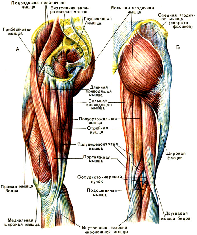
leg muscles
The muscles of the lower leg allow you to raise and lower the foot. The extrinsic calf muscles lower the foot and flex the sole, while the posterior calf muscles raise the heel and allow you to stand on your toes. The foot contains eleven muscles of various sizes and shapes. They are responsible for the movements of the fingers, the separation of the foot from the surface (in total, thirty-eight different muscles of the body are used to perform this function).
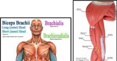
The muscular structure of the arms is very numerous. It is very difficult to list all the names, since only the forearm has about twenty beams. It is worth highlighting that there are two types of muscle fibers - extensors behind the bones, flexors - in front. They envelop the entire structure of the upper limb and allow it to fulfill its functional purpose. The muscles of the hand are divided into tenars, gotenars and middle ones.
Circulation and nerve fibers
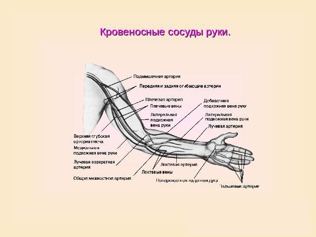
This part of the structure provides the limbs with blood supply (oxygen and micronutrient supply) and innervation (impulses to and from). The upper limbs are fed by the subclavian artery, which then passes into the axillary and brachial. And then the branches connect and flow into the ulnar artery, from which there is a branching further along the arm and hand.
The venous network has a similar structure, and there are also subcutaneous vessels from both parts of the arm. The nervous system begins in the shoulder area. And then the branching passes through the nerve columns - axillary, musculocutaneous, radial, median fibers and ulnar.
The blood circulation of the legs also passes in three directions - arteries, veins and capillaries. The latter are small vessels located close to the surface of the skin. They receive blood from eight large leg veins, which in turn connect to the femoral vein. Arteries pass through the entire leg - femoral, tibial (anterior and posterior), under the knee, dorsal for blood supply to the foot, lateral, medial (the last two on the sole).
The nerve channels in the leg start from the lumbar region and move to the thigh nerve. The innervation of the lower leg is carried out by the fiber of the perineum, tibial and subcutaneous. Three nerves are responsible for the foot - medial, lateral and gastrocnemius.
All nerve channels are interconnected and impulses pass from one to another. Such a system provokes referred pain. When the localization of the injury is in one place, and the pain syndrome spreads to several parts of the limb. Or a problem in the spine, but the leg hurts.
In the video you will see the detailed structure of the muscles of the limb.